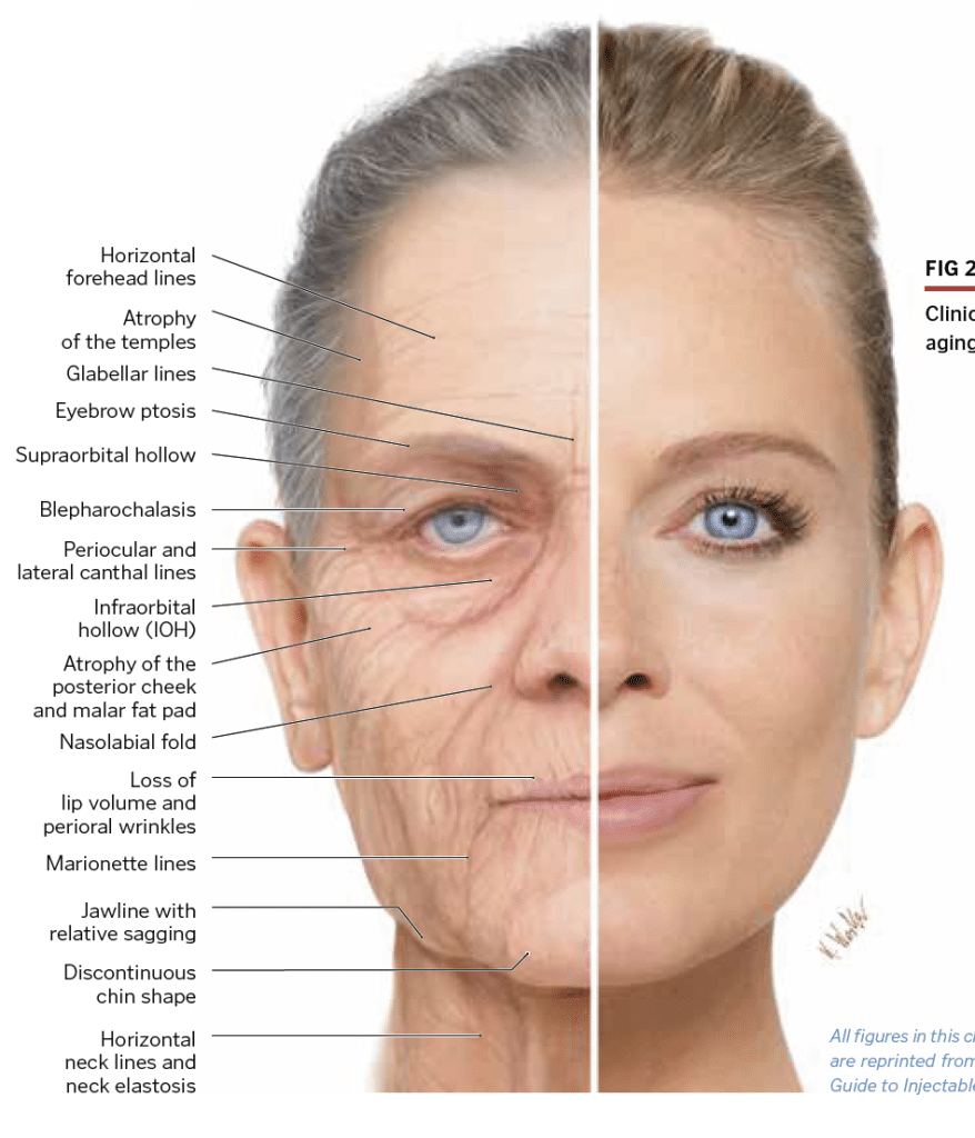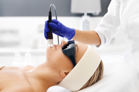
Aging skin is an inevitable process that occurs as we gradually get older. Several factors have been associated with this process which include both genetic and environmental factors.Exposure to sun, pollution, and various chemicals have been known to cause skin and/or DNA damage speeding the aging process.A number of changes to the skin may occur as a result including skin atrophy, telangiectacia, fine and deep wrinkles, yellowing (solar elastosis) and dyspigmentation.Furthermore, a number of additional physical/environment factors including poor diet, lack of exercise, caffeine intake, smoking and drug use are additional factors known to speed the aging process.
Facial anatomy
Facial anatomy remains one of the fundamental aspects for any facial esthetic regimen and the dentist is well situated as much educational background is provided during dental school. It remains important to understand and review facial anatomy, facial features and facial landmarks to safely implement the various therapies described. The face is comprised of various layers including the skin, connective tissue, subcutaneous fat layers as well as muscles, ligaments and underlying bone. Within this network, an array of arteries, veins and nerves also exist.
As such, the role of the clinician in understanding these facial features, characteristics and the anatomy underlying the face becomes pivotal to minimize and counteract the aging process. The desire to achieve a rejuvenated look has become increasingly popular owing to its generally lower costs associated with treatment (when compared to the 90s for instance) which has made it more frequently accessible to more individuals.
Characteristics of age-related facial anatomy changes
Regardless of how beautiful one’s appearance was in their youth, age-related changes and loss of facial volume and features is inevitable. These are often more pronounced and specific to certain areas. A gradual loss of soft tissue occurs in the upper midface region in conjunction with a downward migration of superficial buccal fat. Consequently, the upside-down triangle associated with a youthful look becomes inversed with a larger proportion of soft tissue drooping below the midface found with aging. Both males and females tend to have different ideal characteristics when assessing overall attractiveness and the rate of aging varies between all individuals based on genetics, environmental factors, sex and ethnicity.
Subcutaneous fat and connective tissue
The subcutaneous fat embedded in the connective tissue of the face acts as a ‘volumizing cushion’ of facial soft tissues. It plays a prominent role protecting the face from external injury but also ensures a continuous supply of essential fluids and nutrients to facial tissues. In the face, areas with high fat compartments are typically well defined and homogeneous in layer. These include the cheeks, nasolabial folds, the glabella, and the jaw-chin region. In older patients, this specific tissue decreases with age with a resulting atrophy typically caused from reduced blood flow. Notice also that a very thin subcutaneous fat layer exists in the area of the temples and forehead, and almost none exists in the periorbital and perioral region. These areas are therefore more prone to wrinkles and folds with aging and are one of the first visible sign of facial aging in individuals.
Blood supply
A prominent and complex blood vascular network exists practically throughout the entire region of the face. The peripheral skin layers receive its blood supply from fine capillary vessels. These small vessels allow adequate diffusion into all blood layers. When injecting into areas of the face, a thorough knowledge regarding the whereabouts of the major blood vessels is crucial. This will avoid potential complications related to intravascular injections, most commonly reported with fillers. Importantly, the anatomy of the facial arteries and veins is important.
Innervation
Along with blood supply to the face, a complex innervation system exists within the face mainly from 2 sources: the trigeminal and the facial nerve. The sensory innervation of the face is provided by the trigeminal nerve. This nerve is divided into 3 branches including the V1 ophthalmic nerve which exists the orbit via the supraorbital foramen and fissure and supplies sensation to the upper part of the face. The V2 maxillary nerve which exists from the infraorbital foramen and innervates the midface. And the V3 mandibular nerve that innervates the mandibular and temporal regions.


